SAHA INSTITUTE OF NUCLEAR PHYSICS
Department of Atomic Energy, Govt. of India
| Synchrotron Facility at Raja Ramanna Centre for Advanced Technology, Indore, MP INDUS-II Grazing Incidence X-ray Scattering (GIXS) Beam Line No.-13 |
| Beamline Overview: The Grazing Incidence X-ray Scattering Beamline (BL-13) has been designed, installed, operated and maintained by Surface Physics and Material Science Division of Saha Institute of Nuclear Physics, Kolkata. The beamline is used for grazing incidence x-ray scattering studies of surfaces and interfaces in ambient, low and high temperature and UHV conditions. Also, in-situ and real-time properties change of materials under different conditions will be studied in the beamline. It covers a wide range of techniques such as: * Powder diffraction * Single crystal and multilayer diffraction (high resolution rocking curves) * X-ray reflectivity and diffuse scattering from multilayer thin film structures * In- and out-of-plane grazing incidence diffraction and diffuse scattering. Results obtained from a combination of these techniques give precise information about crystallinity, lattice mismatch in epitaxial films, interface roughness, thickness and electron density of multilayers and superlattices. |
| Source | 1.5 Tesla bending magnet at port-13 of Indus-2 ring |
| Operational Mode | Monochromatic X-ray (wavelength tunable by Double Crystal Monochromator) |
| Beam Acceptance | 1.5 mrad (Horizontal) 0.2 mrad (Vertical) |
| Focusing optics | Vertical focusing by two cylindrical mirrors and horizontal focusing by sagittal bending of DCM 2nd crystal |
| Focal Spot at sample position |
0.30 mm (Horizontal) X 0.10 mm (Vertical) at 8 keV |
| Flux | 1011 photons /sec/cm2 at 8 keV |
| Spectral Resolution |
|
| Beamline Description: The total length of the beamline, from the tangent point of the ring to the sample position, is about 32 m. The front-end part of the beam port is coupled to the beamline through a water-cooled Be window. The main components of the beamline optics consist of a vertical collimating mirror (VCM), a double crystal monochromator (DCM), and a vertical focusing mirror (VFM). The combination of the two mirrors focuses the beam vertically to the sample or detector position in the diffractometer in the experimental station. The DCM monochromatizes the beam and transmits photons between 5 keV and 20 keV. The second crystal of the DCM is bendable and focuses the beam horizontally to the sample or detector position depending on the requirement of the experiments. A nano-BPM placed just before the sample is used to image the beam as well as the beam profile. An ion-chamber placed before the sample can be used to normalize the incident beam of the synchrotron radiation. |
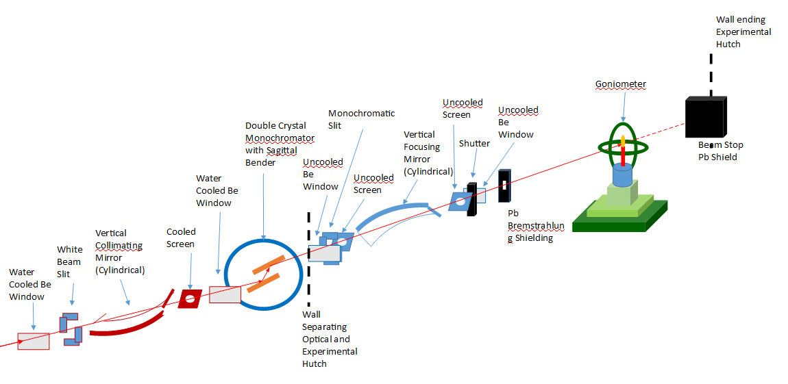
Schematic layout of GIXS (BL-13) beamline (not to scale)
 |
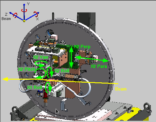 |
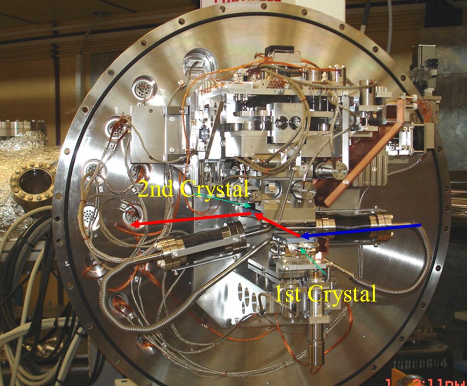
Inside view of the DCM (left) and its schematic showing different motors of the DCM (right)
 |
 |
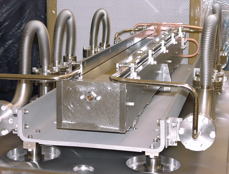
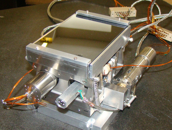
| Inside view of the water cooled VCM (at left) and its schematic showing different motors of the VCM (right) |
|
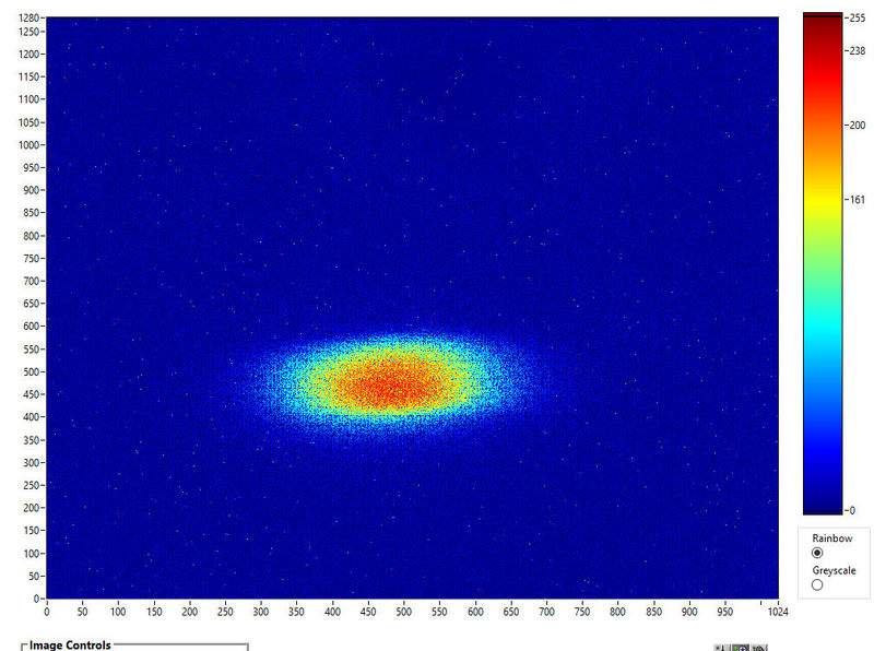 |
| Nano-BPM image of the focused x-ray beam |
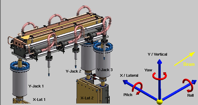
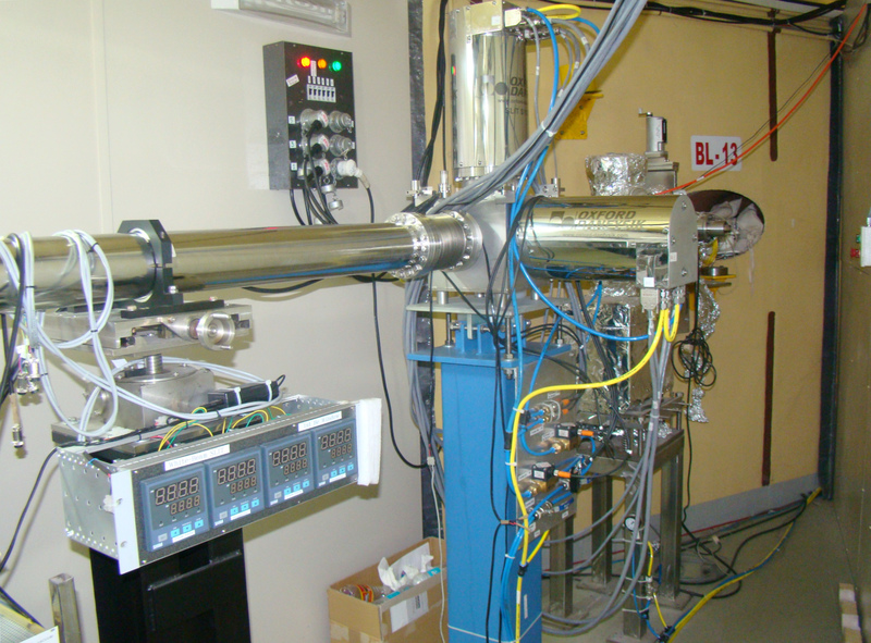
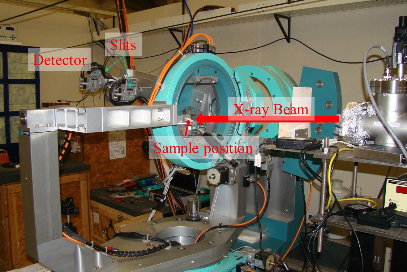 8-circle goniometer of BL-13 |
We use an 8-circle x-ray goniometer for carrying out all our x-ray scattering measurements. A close-cycle helium cryostat can be mounted on the goniometer for doing powder XRD at sample temperature in the range of 10-300 K. A high temperature cell can also be mounted in the goniometer for high temperature measurement under inert ambient or in-vacuum condition (will be available soon). A scintillation detector (C400, Oxford Instruments.) is mounted at the detector arm of the goniometer with crossed slit assembly to count the scattered beam from the sample. For versatility in measurements, we have interfaced the Goniometer with SPEC based data acquisition system. This enables us flexibility to give different combination of coupled motor movements, define the step size, count time etc. |
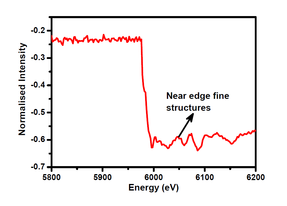
| Recent Results: Energy Calibration: |
|
 |
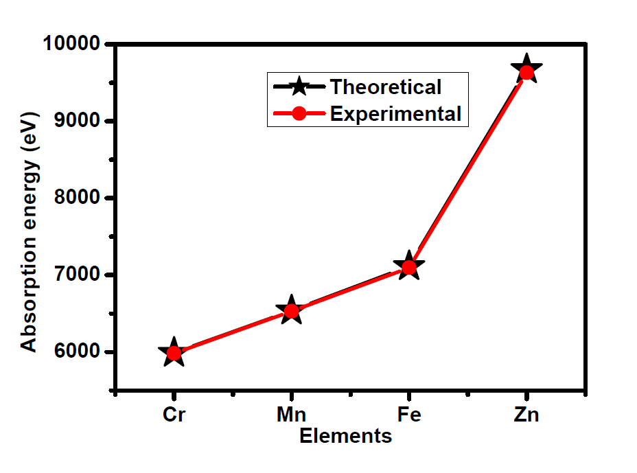 |
| X-ray Absorption Spectra of Cr | Theoretical and experimental absorption edge of elements |
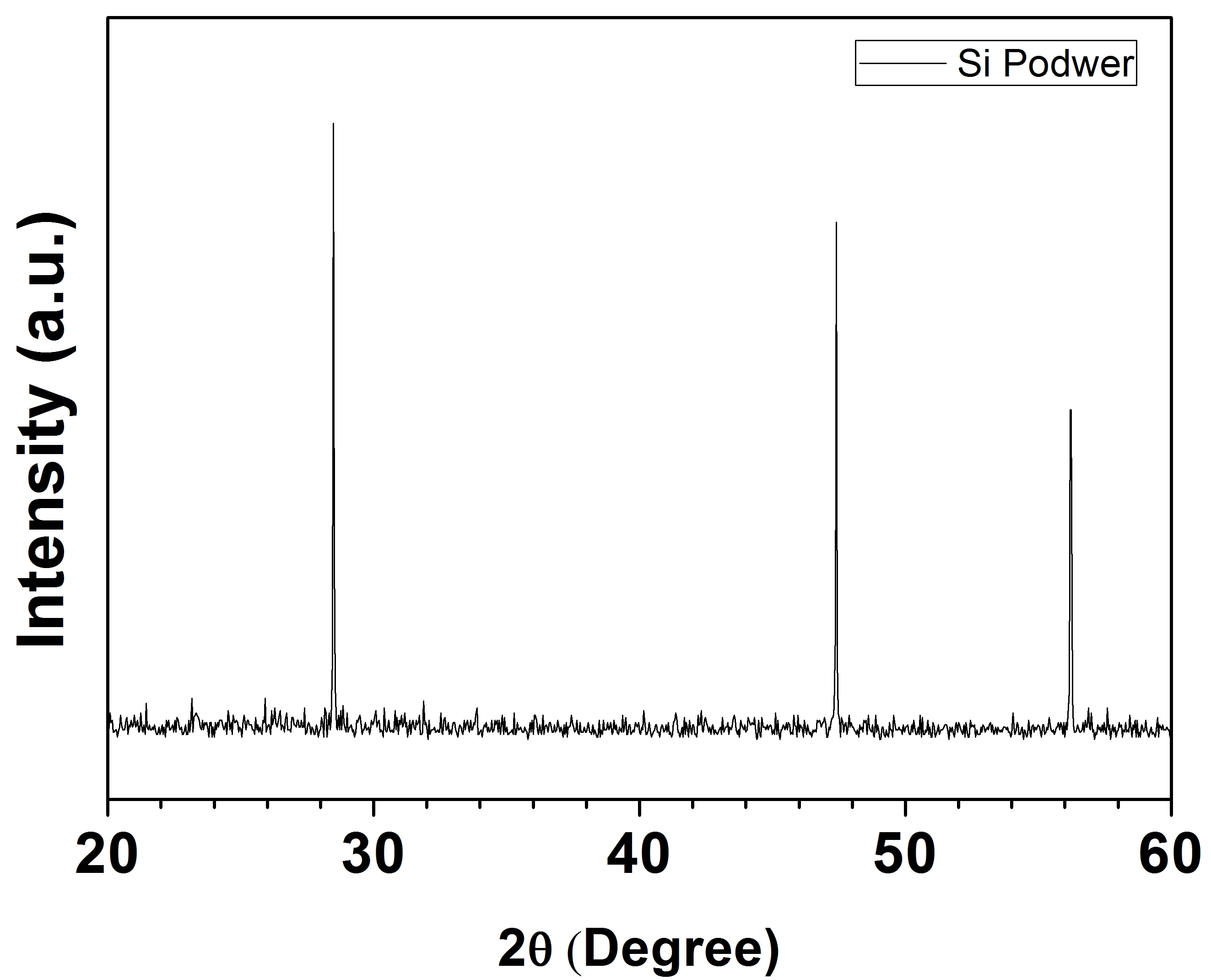 |
| X-ray powder diffraction from standard Si |
 |
||
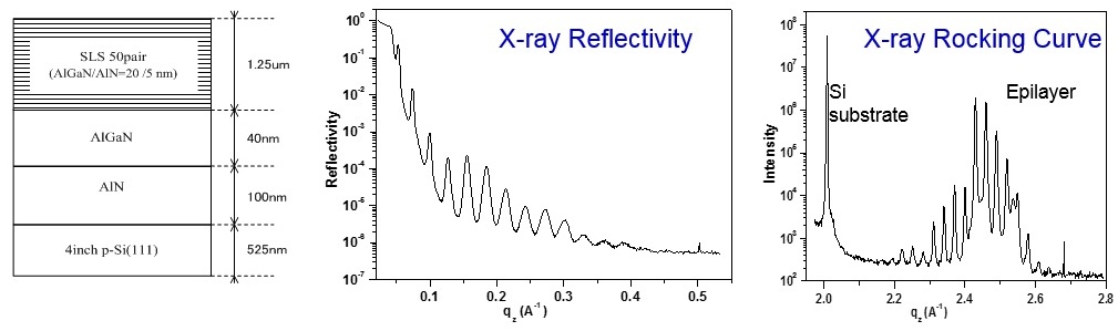
| Schematic of AlN/AlGaN superlattice structure grown on Si |
||
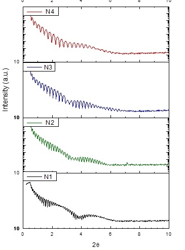 |
| Reflectivity from the niobium oxide thin film on Si substrate at different growth conditions. |
 Official inauguration of BL-13 by Prof. Ajit Kumar Mohanty, Director, SINP and Dr. Prasad A. Naik, Director, RRCAT in December, 2018. |
| INDUS-II Beamline No-13 Phone Number : 0731-244-2513 |
|
| Prof. Satyaban Bhunia, [ Beamline in-charge ] Email: satyaban.bhunia[a]saha.ac.in, Ph: 033-2337-5345 extn 3320 Surface Physics and Material Science Division Saha Institute of Nuclear Physics, Kolkata |
Prof. Mrinmay K. Mukhopadhyay, [ Beamline in-charge ] Email: mrinmay.mukhopadhyay[a]saha.ac.in, Ph: 033-2337-5345 extn 2521 Surface Physics and Material Science Division Saha Institute of Nuclear Physics, Kolkata |
| Mr. Avijit Das Email: avijit.das[a]saha.ac.in, Ph: 033-2337-5345 extn 1117 Surface Physics and Material Science Division Saha Institute of Nuclear Physics, Kolkata |
Dr. Rijul Roychowdhury Research Associate Email: rijul.roychowdhury[a]saha.ac.in Ph ; 0731-244-2513 ( INDUS-II Beamline No-13, RRCAT ) Surface Physics and Material Science Division Saha Institute of Nuclear Physics, Kolkata |
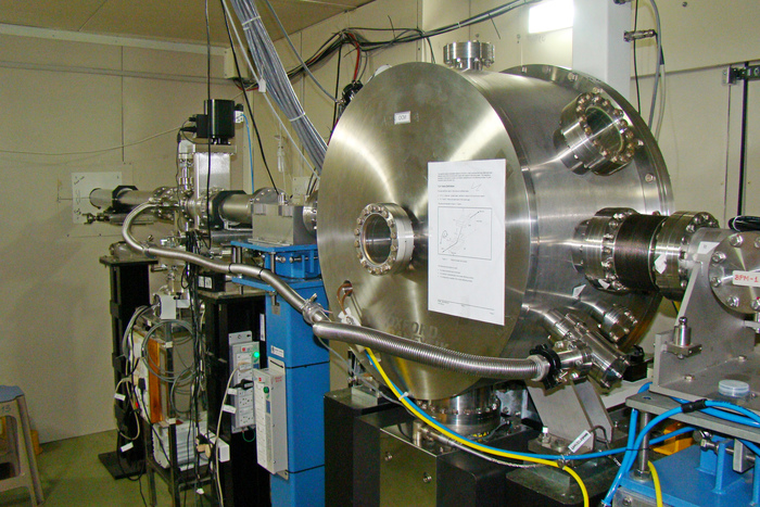
Few Photographs of the beamline and its major components
 |
 |
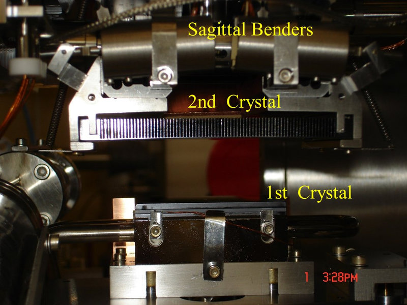
| Inside view of Oxford-Danfysyk Double Crystal Monochromator. | Second Crystal assembly of DCM before installation |
 |
|
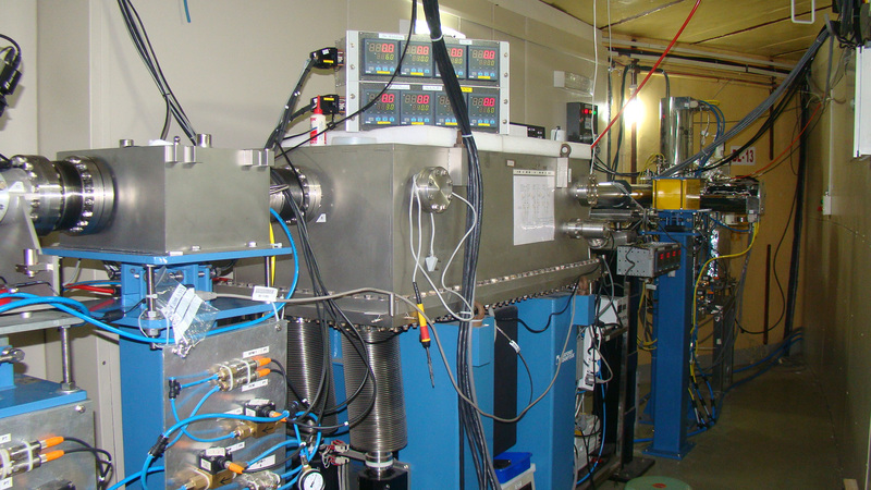
| Part of the optic hutch, where 1st Bremsstrahlung Collimator, Vertically Collimating Mirror and water cooled White Beam Slit is visible outside the Synchrotron Storage Ring wall |
|
 |
 |
| water cooled White Beam Slit |
Another view of BL-13 optic hutch. |
| |
|