Facilities
Microscopy
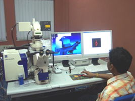
Keeping the demands of the cell biologists in view, the first Confocal Imaging facility (Zeiss LSM510), equipped with live cell imaging attachments, was installed and made operational in 2007. In another three years the system was upgraded to Multi-photon imaging, augmented with a new LSM710 microscope, in vivo FCS and FLIM apparatus. The two systems together churned out beautiful data over the years not only for SINP users, but outsiders as well. In 2012, the facility got a Laser-capture Micro-dissection (LMD) unit. In 2014, a Nikon Super-resolution Microscope, supplemented with a Micro-manipulator was installed. The facility, under one roof would reach its full potential once the in house animal facility is in place. The division is procuring a TIRF microscope in near future. Apart from this divisional facility the institute has TEM, SEM, AFM and several other facilities to do cutting edge research on surface biophysics and physics.
