


Dr. Biswarup Satpati
| Scientist | ||
| Room No | : | Isotope Bldg |
| Ext. | : | 4215 (office) /3128 (lab) |
| Email id | : | biswarup.satpati[AT]saha.ac.in |
| Division | : | |
| Personal Webpage | ||
Research ****************************************************************************************************
*****************************************************************************************************************************************
Growth of low dimensional metal and semiconducting heterostructures
Understanding the appearance of different anisotropic nanostructures in chemical synthesis
The interaction of energetic ions with solids (nanoparticles)
Scanning/Transmission Electron Microscopy (S/TEM)
In-situ Transmission Electron Microscopy
Electron Energy Loss Spectroscopy (EELS) using HRTEM
Nanopatterning
Few Recent Works
****************************
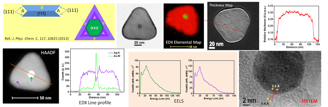
The appearance of different anisotropic one-dimensional (1D) and two-dimensional (2D) gold (Au) core−silver (Ag) shell nanostructures in a single reaction environment. High-resolution transmission electron microscopy (HRTEM) images taken from the core region of a core− shell nanostructure revealed the prominent presence of the hexagonal-shaped gold seed leading to formation of a triangular final particle. The HRTEM studies provide the direct experimental evidence of the “silver halide” model proposed by Sigmund et al. (Lofton, C.; Sigmund, W. Adv. Funct. Mater. 2005, 15, 1197−1208) to explain the kinetic growth mechanism behind their formation. It is important that this information cannot be identified from a single composite nanoparticle due to the lack of atomic number (Z) contrast difference. We have studied energy-dispersive X-ray line profile spectra and elemental mapping using the high-angle annular dark-field scanning/transmission electron microscopy (STEMHAADF) technique corresponding to the Au-M and Ag-K energies from the similar core−shell structures. This confirms the composition of the core to be made of gold and that of the shell of silver. The line profile along the relative thickness map of a hexagonal and triangular nanoplate obtained using energy-filtered TEM indicates the formation of nearly uniform 2D structures. The nearly equal thicknesses of the core and outside shell of a core−shell nanoplate measured using electron energy loss spectroscopy in STEM mode also confirm the 2D growth of a gold seed forming a triangular core−shell nanoplate.
See Tanmay Ghosh and Biswarup Satpati*, J. Phys. Chem. C 2013, 117, 10825−10833 for details
---------------------------------------------------------------------------------------------------------------------------------
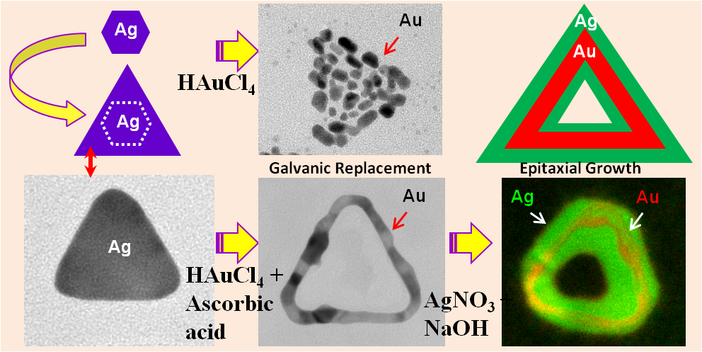
Formation Mechanism of Hexagonal Monometallic Nanopaticles to Triangular Bimetallic Core-Shell Nanorings
Using two-dimensional (2D) triangular silver nanoparticles as templates we first synthesized 2D triangular gold (Au) nanorings via galvanic replacement reaction and then overgrown Ag on Au nanorings via epitaxial growth process. Transmission Electron Microscopy (TEM) and associated techniques were used for in-depth characterization. TEM study reveals that single crystalline Ag nanoparticles led to the formation of continuous Au nanorings with single crystalline walls and the voids spaces which corroborate with the template shapes only when ascorbic acid was added to growth solution. Both the silver nanoplates and gold nanorings are having (111) planes as the basal planes. Subsequently we have synthesized Au-core — Ag-shell nanorings using previously synthesized Au nanorings as template. Energy dispersive X-ray (EDX) line profile spectra and imaging along with high-angle annular dark field scanning/ transmission electron microscopy (STEM-HAADF) techniques were used extensively for compositional study in addition to energy filtered TEM (EFTEM) imaging.
See Tanmay Ghosh, Biswarup Satpati*, Dulal Senapati, Journal of Materials Chemistry C, 2014, 2, 2439 for details----------------------------------------------------------------------------------------------------------------------------------Dendritic silver nanostructures were prepared by a simple dip-and-rinse galvanic displacement reaction directly on germanium surfaces. The formation and evolution of these dendrites were investigated using scanning electron microscopy (SEM), high-resolution transmission electron microscopy (HRTEM) and energy-dispersive X-ray spectroscopy (EDX). The present results clearly show a new kind of heteroepitaxy, where large lattice mismatch between silver and germanium is accommodated at the interface by the formation of low-energy asymmetric tilt boundaries. Over grown sample relax strain by introducing crystal defects. Additionally, employing cathodoluminescence (CL) spectroscopy and imaging in a field emission gun scanning electron microscope (FEG-SEM), we provide information of surface plasmon assisted photon emission from a stack of Ag hexagon nanostructure. Surface enhanced Raman scattering (SERS) study shows the suitability of such Ag nanodendrite structures to be used as SERS active substrate.
See Tanmay Ghosh, Pabitra Das, Tapas Kumar Chini, Tapas Ghosh and Biswarup Satpati, Physical Chemistry Chemical Physics, 2014, 16, 16730 for details
----------------------------------------------------------------------------------------------------------------------------------Study of inelastic mean free path of metal nanostructures using energy filtered transmission electron microscopy imagingWe report a simple method for measuring the inelastic mean free path of nanostructures of known geometry using energy filtered transmission electron microscopy imaging. The mean free path of inelastic electrons was measured by using systems having known symmetry, such as cylindrical or cubic, combined with Poisson statistics without employing the knowledge of microscope parameters, namely the convergence angle and the collection angle.Having inherent symmetry of such systems, their absolute thickness can be measured from their two-dimensional projection images. We have calculated mean free path of inelastic scattering of electrons in gold, silver and nickel doing case study research by employing gold nanorod, silver nanocube and nickel nanorod lying on a carbon-coated TEM grid at two different electron energies (viz. 200 and 300 keV) following this alternative approach. Results obtained using such alternative approach were verified using microscope parameters.
See Tanmay Ghosh, M. Bardhan, M. Bhattacharya and Biswarup Satpati*, Journal of Microscopy Vol. 258, Issue 3 2015, pp. 253–258 for details
----------------------------------------------------------------------------------------------------------------------------------
Room Temperature Ion-Beam-Induced Recrystallization and Large Scale Nanopatterning
We have studied ion-induced effects in the near-surface region of two eutectic systems. Gold and Silver nanodots on Silicon (100) substrate were prepared by thermal evaporation under high vacuum condition at room temperature (RT) and irradiated with 1.5 MeV Au2+ ions at flux ≈1.25×10¹¹ ions cm−2 s−1 also at RT. These samples were characterized using cross-sectional transmission electron microscopy (XTEM) and associated techniques. We have observed that gold act as catalysis in the recrystallization process of ion-beam-induced amorphous Si at room temperature and also large mass transport up to a distance of about 60 nm into the substrate. Mass transport is much beyond the size (∼6–20 nm) of these Au nanodots. Ag nanoparticles with diameter 15–45 nm are half-way embedded into the Si substrate and does not stimulate in recrystallization. In case of Au nanoparticles upon ion irradiation, mixed phase formed only when the local composition and transient temperature during irradiation is sufficient to cause mixing in accordance with the Au–Si stable phase diagram. Spectroscopic imaging in the scanning TEM using spatially resolved electron energy loss spectroscopy provides one of the few ways to measure the real-space nanoscale mixing.
See: T. Ghosh and B. Satpati, Journal of Nanoscience and Nanotechnology, Volume 15, Number 2, February 2015, pp. 1162-1170(9)----------------------------------------------------------------------------------------------------------------------------------
Synthesis of nano-crystalline materials in open-air laboratory: a case study of Tobacco
The work deals with synthesis of nano-crystalline materials in open-air laboratory and in-depth investigation of the tobacco sample of one branded cigarette and its ash using high-resolution transmission electron microscopy and associated techniques. It exhibits the presence of nanocrystals and nanorods of various oxides in cigarette ash. The structure, shape, size and composition of these nanocrystals and nanorods are explored. The energy dispersive X-ray spectra from different regions of the tobacco sample and its ash using high-angle annular dark field scanning/transmission electron microscopy mode are utilized to obtain elemental composition and their relative abundances. For a detailed distribution of different elements in the nanorods, elemental mapping using energyfiltered transmission electron microscopy is also presented. The results highlight the conversion of amorphous constituents of tobacco to nanomaterials on combustion at low temperatures, thus mixing up in the atmosphere.
See: B. Satpati, A. Bhattacharjee and M. Roy, Journal of Nanoscience and Nanotechnology, Volume 15, Number 2, February 2015, pp. 1362-1367(6)-----------------------------------------------------------------------------------------------------------------------------Annealing Induced Morphology of Silver Nanoparticles on Pyramidal Silicon Surface
Here we reports on a simple and cost-effective process of developing a stable surface-enhanced Raman scattering (SERS) substrate based on silver (Ag) nanoparticles deposited on silicon (Si) surface. Durability is an important issue for preparing SERS active substrate as silver nanostructures are prone to rapid surface oxidation when exposed to ambient conditions, which may result in the loss of the enhancement capabilities in a short period of time. Here, we employ the galvanic displacement method to produce Ag nanoparticles on Si(100) substrate prepatterned with arrays of micropyramids by chemical etching, and subsequently, separate pieces of such substrates were annealed in oxygen and nitrogen environments at 550 °C. Interestingly, while nitrogen-annealed Si substrates were featured by spherical-shaped Ag particles, the oxygen annealed Si substrates were dominated by the formation of triangular shape particles attached with the spherical one. Remarkably, the oxygen-annealed substrate thus produced shows very high SERS enhancement compared to the either unannealed or nitrogen annealed substrate. The hitherto unobserved coexistence of triangular morphology with the spherical one and the gap between the two (source of efficient hot-spots) are the origin of enhanced SERS activity for the oxygen-annealed Ag particle-covered Si substrate as probed by the combined finite-difference time domain (FDTD) simulation and cathodoluminesensce (CL) experiment. As the substrate has already been annealed in an oxygen environment, further probability of oxidation is reduced in the present synthesis protocol that paves the way for making a novel long-lived thermally stable SERS substrate.
Ref: Roy et al., ACS Appl. Mater. Interfaces, 9, (2017) 34405−34415
-----------------------------------------------------------------------------------------------------------------------------
Core-Shell Gold @Silver Nanorods of Varying Length for High SERS Enhancement
We report an effective method to produce surface-enhanced Raman scattering (SERS) substrates with improved enhancement efficiency, high uniformity, greater chemical and physical stability and absolute synthetic reproducibility. Nano bipyramidal (NBP) Au core and Ag shell nanorods (NRs) with variable length were prepared simply by varying the Ag precursor amount during growth and could successfully prepared NRs of length from 200 nm to 1200 nm and studied their formation mechanism and morphology using transmission electron microscopy (TEM) and optical properties by using UV-vis-NIR and Raman spectroscopy. Here we have utilized both the properties of Au and Ag nanoparticles by synthesizing Au@Ag core-shell NRs for high SERS enhancement. NRs have stronger absorption and large scattering cross-section for electromagnetic radiation and variation of aspect ratios of these NRs leads to a broad band surface plasmon tuning in the vis-NIR region. Relative SERS enhancement efficiency of these core-shell gold@silver NRs have been measured by using 4-mercaptobenzoic acid (4-MBA), crystal violet (CV) and 4-mercaptopyridine (4-MPy) as Raman tag and the obtained SERS results show their huge enhancement (~1010 for 4-MBA) which was substantiated through finite-difference time-domain (FDTD) computer simulations. Our work show superior SERS enhancement compared to the previously reported result for pure Au and Ag NRs and bimetallic nanorods with Au nanorod as core and Ag shell having aspect ratio in the same range.Ref: Bhattacjarjee et al., ACS Appl. Nano Mater., 2018, 1, 10, 5589-5600---------------------------------------------------------------------
Substrate decomposition in galvanic displacement reaction: Contrast between gold and silver nanoparticle formationWe have investigated substrate decomposition during formation of silver and gold nanoparticlesin galvanic displacement reaction on germanium surfaces. Silver and gold nanoparticles were synthesized by electroless deposition on sputter coated germanium thin film (∼ 200 nm) grown initially on silicon substrate. The naoparticles formation and the substrate corrosion were studied using scanning transmission electron microscopy (STEM) and the energy dispersive X-ray (EDX) spectroscopy.
Tapas Ghosh, D. Kabiraj and B. Satpati, AIP Conf. Proc. 1665, 080040 (2015); http://dx.doi.org/10.1063/1.4917944
---------------------------------------------------------------------------------------------------------------------------------
*******************************************************************************************************************************************
*******************************************************************************************************************************************My Photography
**********************

.png)

.png)
.png)
.png)
.png)

