Underlying ripple structure in ion beam induced Si wafers
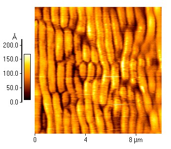 The formation of periodic ripple or wave-like pattern with a spatial periodicity varying from nano to micro range on obliquely ion-bombarded solid surfaces has become a topic of intense research in the context of fabrication of nanoscale textured materials, such as templates for growing nanowires, nanorods or nanodots. Ion-induced ripples are thought to be produced by interplay between a roughening process caused by the ion beam erosion (sputtering) of surface and a smoothening process caused by thermal or ion-induced surface diffusion. The balance between positive surface diffusion and negative surface tension develops instability along the projection of the ion beam and also perpendicular to it, leading to ripple formation in both directions. However, experimentally observed ripple structure has the direction for which the growth rate is largest and is generally along the projection of the ion beam consistent with current theoretical models.
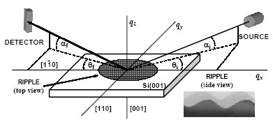
It is an established fact that monocrystalline semiconductors may be rendered amorphous easily compared to crystalline metals under room temperature keV energy heavy ion (such as Ar) impact. However, our conventional wisdom on ion-solid interaction does not answer the question whether the amorphous/crystalline interface underneath the top surface maintain its planarity when the semiconductor surface under oblique ion bombardment transforms its initial flat geometry to a corrugated one. This is a relevant question on accounting the extent of the disordered zone responsible in the smoothening mechanism by viscous relaxation assumed in some recent models6 of ripple formation in semiconductors. Interestingly, quite recently cross-sectional transmission electron microscopy (XTEM) study on obliquely incident (50-120) keV Ar+ bombarded Si surface shows that crystalline part at the buried amorphous crystalline interface also follows the same sinusoidal ripple pattern as formed in the top amorphous layer. The Ar+ dose employed in this study was about 1018 ions/cm2, an order of magnitude higher than the critical dose (1017 ions/cm2) for ripple formation under similar experimental condition. Although, XTEM can provide unique information about out of plane interfacial structure, it can not give the information about in-plane inter-facial structure that is essential to understand the process of ripple evolution for the Ar-Si combination through the study of top as well as buried damaged layer in the dose range where the ripple initiate to a stage of full growth (dose ~1016-1018 ions/cm2).
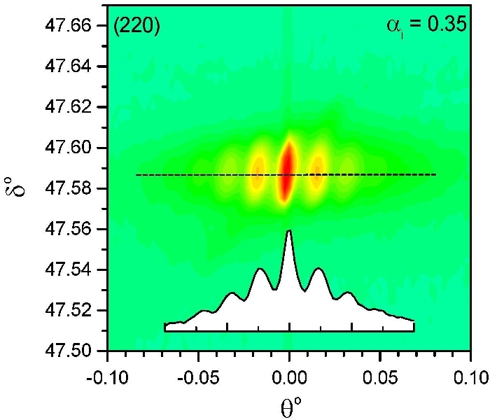
Ion beam induced ripple formation in Si wafers was studied by two complementary surface sensitive techniques, namely atomic force microscopy (AFM) and depth-resolved X-ray grazing incidence diffraction (GID). The formation of ripple structure at high doses (~7x10¹⁷ ions/cm2), starting from initiation at low doses (~1x10¹⁷ ions/cm2) of ion beam is evident from AFM, while that in the buried crystalline region below a partially crystalline top layer is evident from GID study carried out at ESRF. Such ripple structure of crystalline layers in large area formed in the subsurface region of Si wafers is probed for the first time through a nondestructive technique. The GID technique reveals that these periodically modulated wave-like buried crystalline features become highly regular and strongly correlated as one increases the Ar ion beam energy from 60 keV to 100 keV. The vertical density profile obtained from the analysis of Vineyard profile shows that the density in the upper top part of ripples is decreased to about 15 % of the crystalline density. The partially crystalline top layer at low dose, transforms to a completely amorphous layer for high doses and the top morphology was found to be conformal with the underlying crystalline ripple [published in Phys. Rev. B 70, 121307(R) (2004) and also in ESRF Highlights 2004].
From the study of the in-plane rotation of the samples with respect to the ion-beam, it appears that the ripple vector always follows the direction of ion beam. Ripples still appear for the angle 15o, but nearly disappear for the angle 30o, 45o. From the study of the out-of-plane rotation of the samples with respect to the ion-beam, optimum grating seems to be appear for the angle 65o. In case of optimum grating side peaks appear always in the crystalline part and for non-optimum, it appears even in deeper parts. For optimum grating, we find inclined crystal truncation rods, indicating the appearance of side facets. The value of the angle is in the range of expectation. Side facets seems to be changing with depth.
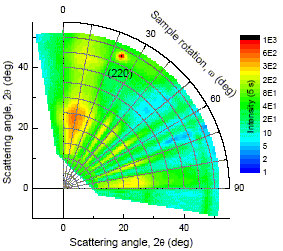
Using grazing-incidence x-ray scattering technique we have investigated the evolution of the damage profile of the transition layer between the ion-induced ripple-like pattern on top surface and the ripples at buried crystalline interface in silicon created after irradiation with 60 keV Ar+ ions under 60o. The transition layer consists in a defect-rich crystalline part and a complete amorphous part. The crystalline regions are highly strained but relaxed for low dose and high dose irradiation, respectively. The appearance of texture in both cases shows that the damage of the initial crystalline structure by the ion bombardment takes place along particular crystallographic directions [published in Appl. Phys. Lett. 89, 231900 (2006)].
Si(001) surface, modified using low energy (5 keV) Ar+ ion-beam, is studied by scanning tunneling microscopy (STM) under ultrahigh vacuum conditions. The evolution of surface for ion bombardment in presence of temperature is observed through STM images and is characterized by height-height correlation function. It has been observed that the temperature plays an important role along with the dose for the control of the surface evolution, especially, post annealing helps to form nanoscale ripple-like structures at relatively low temperature. The results demonstrate that ion-beam induced and thermal diffusions cannot be treated as additive processes and the observed enhancement of surface diffusion requires lowering of activation energy that arises due to creation of ion-beam induced vacant regions [published in J. Phys. D 41, 055306 (2008)].
|

